What Specific Type Of Unipolar Neuron Is Shown In The Diagram Labeled B
What specific type of unipolar neuron is shown in the diagram labeled b. What specific type of unipolar neuron is shown in the.
 Labeled Neuron Diagram Science Trends
1221 contrast the histological characteristics and the functions of neurons and neuroglia.

What specific type of unipolar neuron is shown in the diagram labeled b. What specific type of unipolar neuron is shown in diagram labeled b. Type i cutaneous mechanoreceptor merkel disc what specific type of unipolar neuron is shown in the diagram labeled b. A axonb cell bodyc dendrited membrane answer.
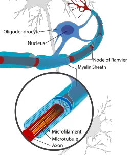
The cell body also known as the soma is the part of. This structure electrically insulates the axon of a neuron to increase the speed of nerve impulse conduction. E sympathetic preganglionic neuron.
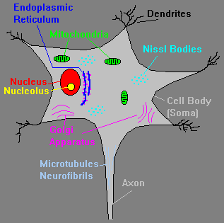
Easy learning objective 1. A type i cutaneous mechanoreceptor merkel disc b corpuscle of touch meissner corpuscle c lamellated pacinian corpuscle d nociceptor e purkinje cell. Which part of the neuron is responsible for manufacturing proteins.
A type i cutaneous mechanoreceptor merkel disc b corpuscle of touch meissner corpuscle c lamellated pacinian corpuscle d nociceptor e purkinje cell. A merkel disc b meissner corpuscle c pacinian corpuscle d nociceptor e purkinje cell ans. Merkel disc which neuroglial cell is located mainly in gray matter and helps maintain the appropriate chemical environment for generation of nerve impulses.
D parasympathetic postganglionic neuron. A type i cutaneous mechanoreceptor merkel disc b corpuscle of touch meissner corpuscle c lamellated pacinian corpuscle d nociceptor e purkinje cell. What specific type of unipolar neuron is shown in the diagram labeled c.
What specific type of unipolar neuron is shown in the diagram labeled b. Medium learning objective 1. 68 which of the labeled cells in the figure is not a neuroglial cell.
B parasympathetic preganglionic neuron. A gangliab bipolarc rodsd cones answer. 122 describe the structures and functions of neurons and neuroglia and distinguish between white matter and gray matter.
The structure labeled 3 in the diagram is a a somatic motor neuron. Which type of receptor cell is associated with seeing colors. What specific type of unipolar neuron is shown in the diagram labeled b what specific type of unipolar neuron is shown in the diagram labeled b nervous system.
What specific type of unipolar neuron is shown in the diagram labeled b photos. 122 describe the structures and functions of neurons and neuroglia and distinguish between white matter and gray matter.
 Morphological And Functional Diversity Of First Order
![]() Neurotransmitters Types Functions And Disorders Kenhub
Neurotransmitters Types Functions And Disorders Kenhub
 Anatomy 102 Chapter 12 Study Guide Diagram Quizlet
 Rabconnectin 3a Is Required For The Morphological Maturation
 Labeled Neuron Diagram Science Trends
 Nerve Tissue The Nervous System Junqueira S Basic
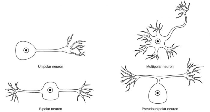
 Unipolar Neuron Wikipedia
The Nervous System
 Introduction From Neurons To The Mind Springerlink
 What Is The Structural Classification Of The Neuron Labeled
 The Kruppel Like Factor Dar1 Determines Multipolar Neuron
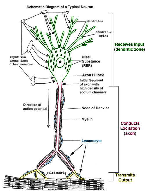
 Nerve Cells Neurons Structure Function Adaptations
 Nerve Tissue The Nervous System Junqueira S Basic
 Robo1 And 2 Repellent Receptors Cooperate To Guide Facial

 The Lpa Lpa4 Axis Is Required For Establishment Of Bipolar
 How Does Migraine Surgery Work Springerlink
 Nerve Tissue The Nervous System Junqueira S Basic
 Neuroscience For Kids Cells Of The Nervous System
What Is The Structural Classification Of The Neuron Labeled
 Full Text Dexmedetomidine Attenuates The Neurotoxicity Of
 Neural Circuitry Of A Polycystin Mediated Hydrodynamic
Belum ada Komentar untuk "What Specific Type Of Unipolar Neuron Is Shown In The Diagram Labeled B"
Posting Komentar