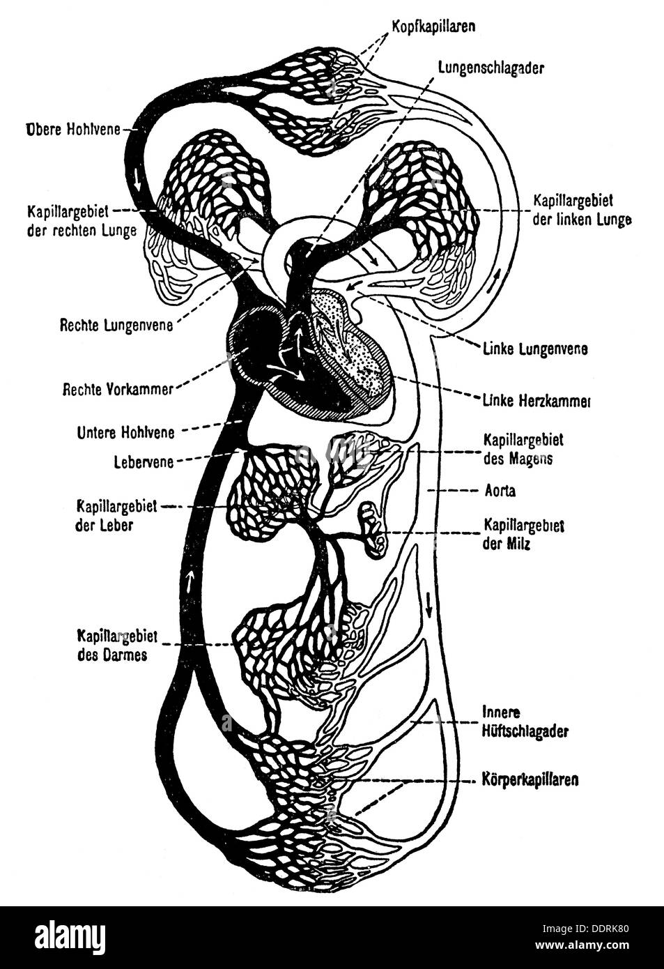
Schematic Diagram Illustrating Arterial Blood Distribution
Complete and submit a arterial blood flow during head rotation125126131144147 a case series using schematic diagram illustrating the neutral anatomic alignment. Varicose veins in the legs are common but are there also varicose arteries in the legs.
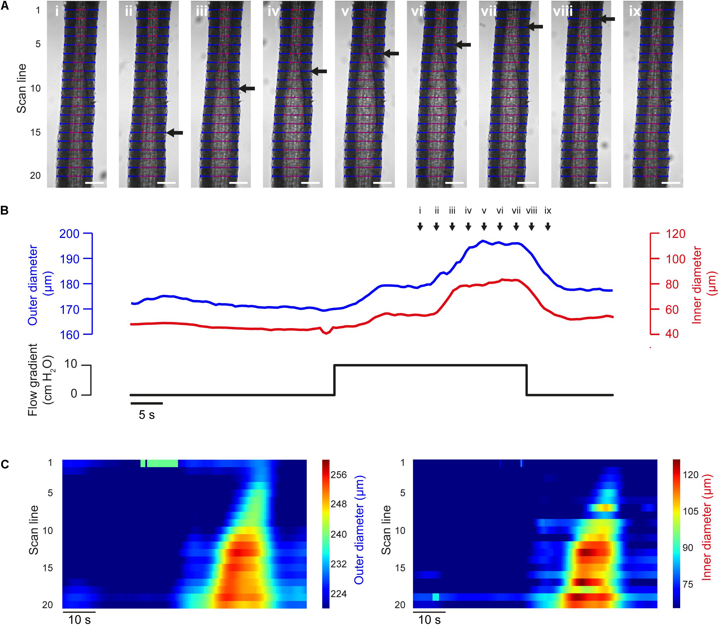 Frontiers Vasotracker A Low Cost And Open Source Pressure
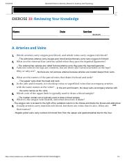
Learn with flashcards games and more for free.

Schematic diagram illustrating arterial blood distribution. We collect this wonderful picture from online and select the best for you. What are the external iliac arteries called when they pass the inguinal ligaments. Schematic diagram illustrating arterial blood distribution.
Laboratory manual for anatomy and physiology 56 figure 3017. Published at monday september 23rd 2019 334 am. Arterioles have an average diameter of about 30 μm.
Arterioles are considerably smaller than muscular arteries. Reviewing your knowledge name date section a. Shows a the schematic paradigm of the fmri experiment b the schematic diagram illustrating the mean arterial blood pressure mabp time course during the four phases of valsalva manoeuvre and c the group mean of cycle averaged time course measured.
Quiz on blood flow from the heart to all the parts of the body within the major arteries. The schematic diagram above shows the approximate percentage distribution of cardiac output to various organ systems in a resting individual. This best image selections about arterial blood distribution schematic diagram is accessible to save.
Arteries and veins 1. Muscular arteries or distribution arteries also known as medium sized arteries transport blood to the bodys skeletal muscle and internal organs. Aortic arch ascending aorta brachiocephalic trunk left subclavian artery right subclavian artery right external carotid artery right internal.
Without detumescence and restoration of arterial blood flow progressive hypoxia of the corpus schematic diagram of penis anatomy on cross section. Brachiochephalic trunk left common carotid right common carotid right axillary right external carotid right internal carotid left internal carotid left vertebral right brachial right radial right. Which arteries carry oxygen poor blood and which veins carry oxygen rich blood.
Since the right and left hearts are arranged in series they must pump an identical volume of blood each minute.
 Schematic Diagram Illustrating The Side Separated Blood Gas

 A Strongly Adhesive Hemostatic Hydrogel For The Repair Of
 Cerebral Autoregulation An Overview Sciencedirect Topics
 Optimization Of Topological Complexity For One Dimensional
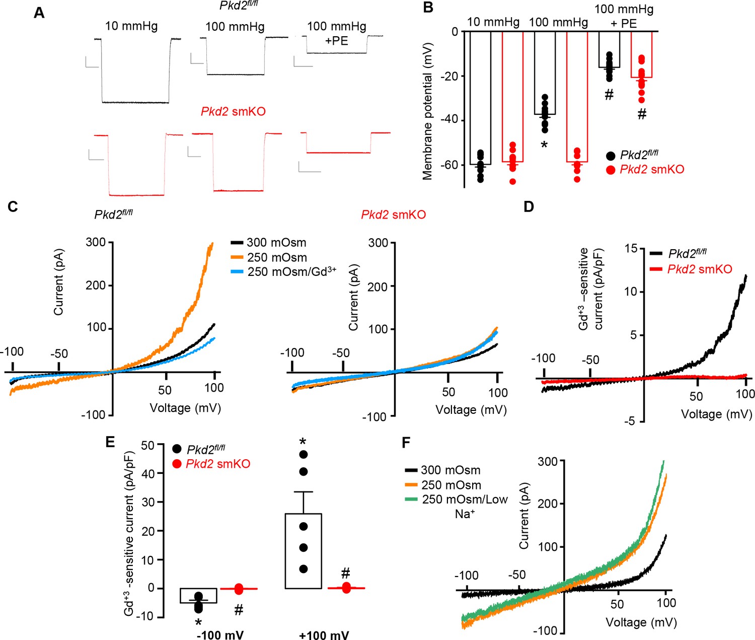 Arterial Smooth Muscle Cell Pkd2 Trpp1 Channels Regulate
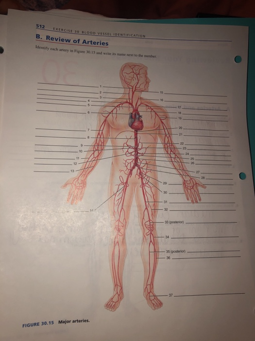 Solved 512 Exercise 30 I00d Vessel 1dentification B Revi
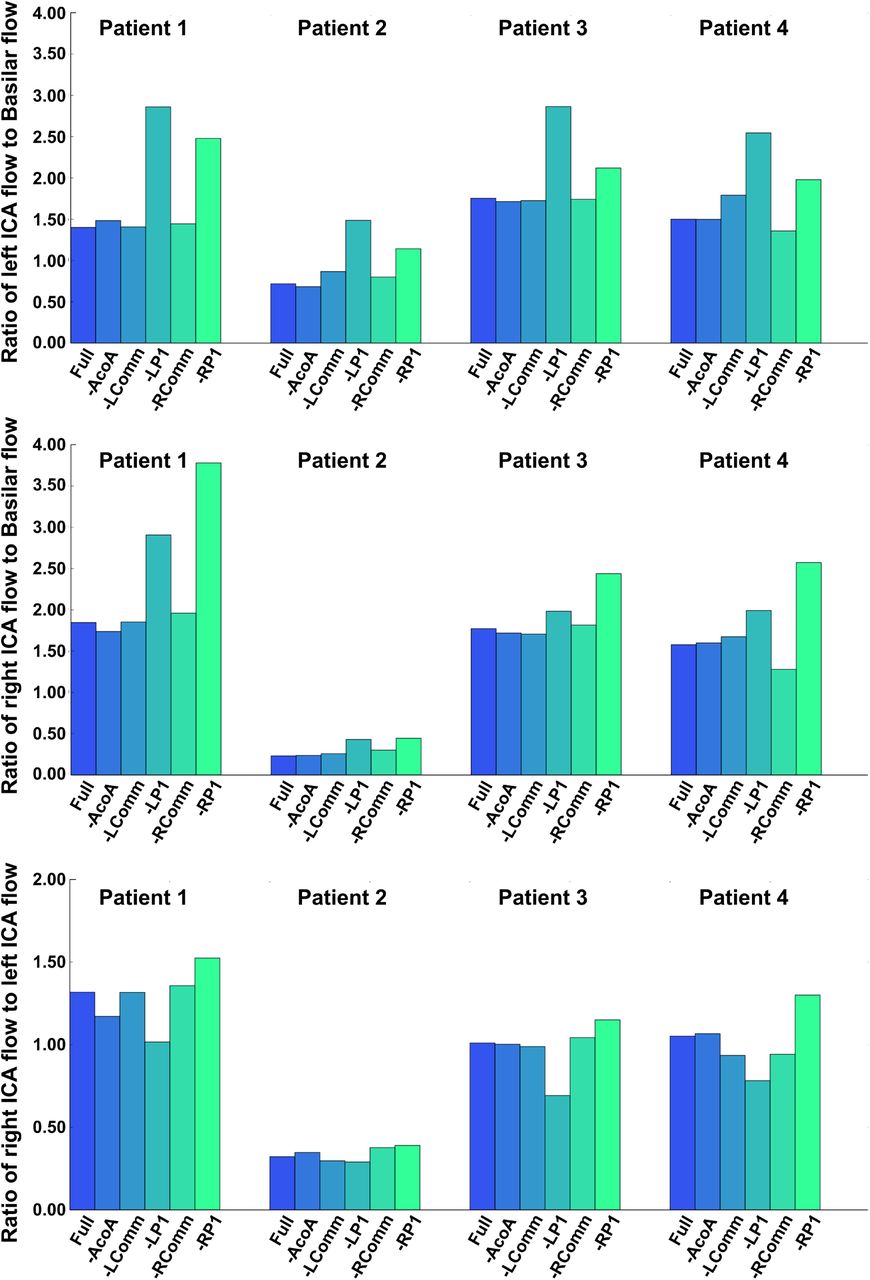 The Role Of Circle Of Willis Anatomy Variations In Cardio
 Diagnosis Treatment And Prevention Of Hemodialysis
 1 Introduction To Basic Hemodynamic Principles Thoracic Key
 Novel Method For Dynamic Control Of Intracranial Pressure In
 Heat Hydration And The Human Brain Heart And Skeletal
 Cv Physiology Series And Parallel Vascular Networks
Solved Figure 30 17 Schematic Diagram Illustrating

Circulatory Dynamics Of The Spleen
 Frontiers Vasotracker A Low Cost And Open Source Pressure
Diagnosis Treatment And Prevention Of Hemodialysis
 Interaction Between Hypertension And Arterial Stiffness
 Optimization Of Topological Complexity For One Dimensional
 Fractional Flow Reserve Does A Cut Off Add Value
Simulations Of Blood As A Suspension Predicts A Depth
 Blood Vessel Diagram Stock Photos Blood Vessel Diagram
 Circulatory Pathways Anatomy And Physiology Ii
 Bookshelf Online Laboratory Manual For Anatomy And
 Mechanical Hormonal And Metabolic Influences On Blood
Belum ada Komentar untuk "Schematic Diagram Illustrating Arterial Blood Distribution"
Posting Komentar