In The Figure Which Diagram Of A Cell Wall Is A Gram Negative Cell Wall
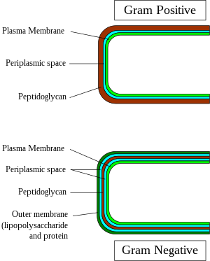
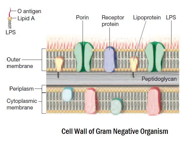
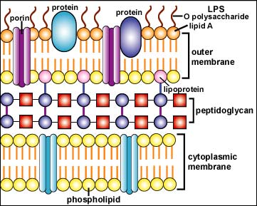
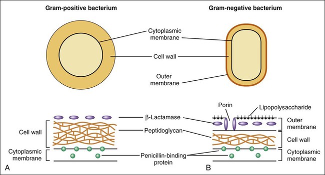
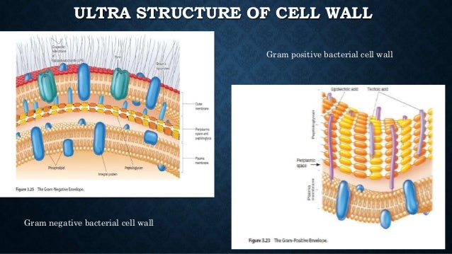
The peptidoglycan portion of the gram negative cell wall is generally 2 3 nanometers nm thick and contains just 2 3 layers of peptidoglycan figure 1c. Gram positive bacteria do not have an outer cell membrane found in gram negative bacteria.
 Compare Contrast This Is A Great Diagram Of The General
In figure 43 which diagram of a cell wall is a gram negative cell wall.

In the figure which diagram of a cell wall is a gram negative cell wall. B it is sensitive to lysozyme. 10 differences between cell wall of gram positive and gram negative bacteria gram staining is a special technique which is used to stain bacteria. The cell wall of gram positive bacteria is high in peptidoglycan which is responsible for retaining the crystal violet dye.
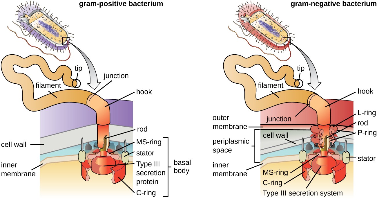
The gram negative bacteria include the model organism escherichia c. Its rigid structure gives the bacterial cell shape surrounds the plasma membrane and provides. C it protects the cell in a hypertonic environment.
Gram negative cell walls. The cell walls of gram negative bacteria are more complex than that of gram positive bacteria with more ingredients overall. Chemically gram stain is a weakly alkaline solution of crystal violet or gentian.
Coli as a single layered cylindrical spring network as shown eg in fig. E it is sensitive to penicillin. 1 and in expanded view in fig.
D it contains teichoic acids. 2 each of the following statements concerning the gram positive cell wall is true except a it maintains the shape of the cell. Gram negative bacteria are bacteria that do not retain the crystal violet stain used in the gram staining method of bacterial differentiation.
This technique was developed by christian gram in 1884. C both a and b in figure 43 which diagram of a cell wall is decolorized by acetone alcohol. The stain stain used in gram staining is called gram stain.
We model the peptidoglycan cell wall of a gram negative rod shaped bacterium such as e. Spring model of gram negative cell wall. Gram negative bacteria are found everywhere in virtually all environments on earth that support life.
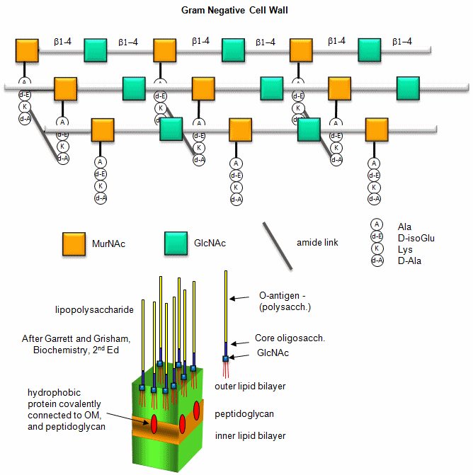
This is due to the difference in the structure of their bacterial cell wall. The gram negative cell wall is composed of a thin inner layer of peptidoglycan and an outer membrane consisting of molecules of phospholipids lipopolysaccharides lps lipoproteins and sutface proteins. In figure 43 which diagram of a cell wall is a gram negative cell wall smaller gram negative in figure 43 which diagram of a cell wall is a toxic cell wall.
They are characterized by their cell envelopes which are composed of a thin peptidoglycan cell wall sandwiched between an inner cytoplasmic cell membrane and a bacterial outer membrane. They do contain peptidoglycan as well although only a couple of layers representing 5 10 of the total cell wall. Peptidoglycan pep tid o gly can is a molecule found only in the cell walls of bacteria.
The lipopolysaccharide consists of lipid a and o polysaccharide. In figure 43 which diagram of a cell wall has a wall that protects against osmotic lysis.
 Surface Proteins Of Gram Positive Bacteria And Mechanisms Of
 A Tour Of The Cell 3 The Organisation Of Prokaryotic Cells
 Schematic Structure Of Gram Positive And Gram Negative Cell
 Bacterial Cell Wall Structure Composition And Types
 Gram Positive Bacteria Wikipedia


Gram Staining Gram Positive And Gram Negative Differences
 3 3 Unique Characteristics Of Prokaryotic Cells
 In Figure 43 Which Diagram Of A Cell Wall Is A Gram Negative

 Diagram Demonstrating Of The Cell Wall Structure Of A
 Cell Wall Definition Function Structure Biology
 Bacterial Cell Walls Structure Function Types Video
 Differences Between Gram Positive And Gram Negative Bacteria
 Structure Of Bacterial Cells Review Of Medical
 Unique Characteristics Of Prokaryotic Cells Microbiology
 2 3b The Gram Negative Cell Wall Biology Libretexts
 Determining The Bacterial Cell Biology Of Planctomycetes
 A Diagram Of A Gram Negative Cell Wall B Electron
 Bacterial Capsule Wikipedia
 Figure 1 1 From The Study Of Ctp Glycerol 3 Phosphate
 Inhibitors Of Bacterial Cell Wall Synthesis Basicmedical Key
 2743 Exam1 Ch04 Biological Sciences 2743 With Sridhar At
 B2 Cell Walls Biology Libretexts

 Bacterial Cell Wall Synthesis
 Pathogen Recognition And Innate Immunity Cell
Belum ada Komentar untuk "In The Figure Which Diagram Of A Cell Wall Is A Gram Negative Cell Wall"
Posting Komentar