Which Protein Subunits Are Depicted In The Diagram
Complex iv the oxidase has 13 in vertebrates. Polypeptide chains generally contain both hydrophobic and hydrophilic residues.
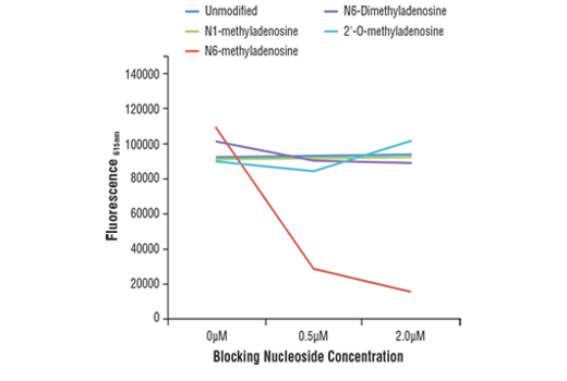 Cst N6 Methyladenosine M6a D9d9w Rabbit Mab
The eukaryotic small ribosomal subunit is the smaller subunit of the eukaryotic 80s ribosomes with the other major component being the large ribosomal subunit.

Which protein subunits are depicted in the diagram. Proteins form by amino acids undergoing condensation reactions in which the amino acids lose one water molecule per reaction in order to attach to one another with a peptide. 8 correct association of the 40s and 60s subunits induces hydrolysis of the gtp bound to elf5. Bacterial ribosomes are composed of two subunits of 30s and 50s sedimentation coefficient in sucrose.
A single amino acid monomer may also be called a residue indicating a repeating unit of a polymer. Many cellular enzymes are composed of subunits. Correct the subunits present depend on whether the ribosome is eukaryotic 40s and 60s or prokaryotic 30s and 50s.
It is structurally and functionally related to the 30s subunit of 70s prokaryotic ribosomes. Each ribosome has two unequal subunits a large and a small subunit. They have combined sedimentation coefficient of 70s.
Elf5 gtp is now bound to elf1a in the a site. The same classes of interactions that contribute to the stability of tertiary protein structure also serve to stabilize the quaternary association of subunits namely ionic bonds hydrogen bonds hydrophobic bonds and disulfide bridges. Three each of alpha and beta and 8 of little c again in vertebrates.
Part c which protein subunits are depicted in the diagram. Then elf5b gdp and elf1a are released. The atp synthase has multiple subunits some of which are present in multiple copies within the same molecule.
Since this cap is present in the diagram the. Select all that apply. Different subunits belonging to the same protein plex different subunits belonging to the same protein plex often exhibit discordant expression levels and evolutionary properties figure s1 a mechanism of covalent substrate binding in the x ray fig 1 structure of the dhakdha plex a ribbon diagram of the dhak dimer the subunits are gray the n terminal.
Much like detergent micelles proteins are most stable when their hydrophobic parts are buried while hydrophilic parts are on the surface exposed to water. The ribosome moves down the mrna in the 5 to 3 direction and synthesizes protein in the direction of amino terminus to carboxyl terminus. Vertebrate complex iii has 11 subunits all different.
Proteins are polymers specifically polypeptides formed from sequences of amino acids the monomers of the polymer. This accurately describes the polarity with which the message is read as well as the direction of protein synthesis. A ribosome has two subunits.
The 7methylguanosine m7g cap is added to the 5 end of the premrnas in eukaryotes. Both subunits contain many proteins and at least one large rrna. 7 the 60s subunit will now combine with the 40s subunit resulting in release of most of the initiation factors.
Ribosomes and protein synthesis. Primary secondary tertiary quaternary. Complex i the most complex has 49 or 50.
Protein structure is the three dimensional arrangement of atoms in an amino acid chain molecule. However the 40s subunit is much larger than the prokaryotic 30s subunit and contains ma. The 40s and 60s names originate from the convention that ribosomal particles are denoted according to their sedimentation coefficients in svedberg units.
 Human Dna Polymerase Delta Is A Pentameric Holoenzyme With A
 Local Frustration Around Enzyme Active Sites Pnas
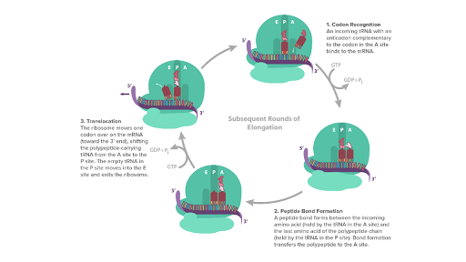
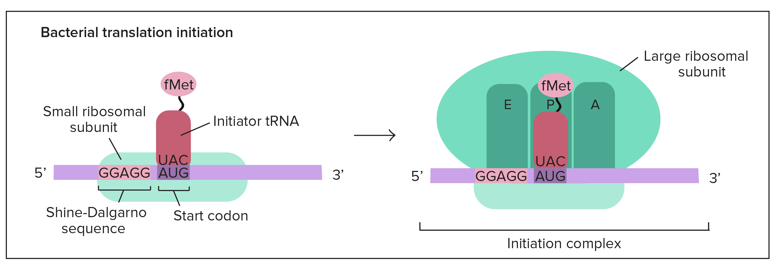
![]() Translation Of Dna Initiation Elongation Termination
Translation Of Dna Initiation Elongation Termination

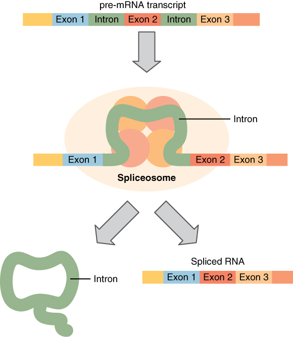
 Storing Genetic Information Biology For Non Majors I
 Rna Protein Interactions Disorder Moonlighting And Junk
 A Potential Architecture Of The Ino80 Complex The 15
 Dynamics Of Ribosomes And Release Factors During Translation
 Rna Protein Interactions Disorder Moonlighting And Junk
Bead Aggregation Assays A Schematic Representation Of
 Cell Surface Protein Identification Dualsystems Biotech Ag
Figure 12 From Cech Single And Double Stranded Dna Primers
The Science Behind G Protein Coupled Receptors Gpcrs And
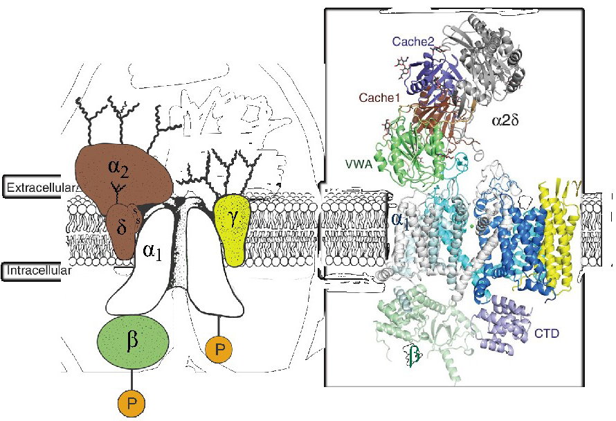 Voltage Gated Calcium Channels Introduction Bps Iuphar

 Fig 7 Microbiology And Molecular Biology Reviews
:max_bytes(150000):strip_icc()/what-are-the-parts-of-nucleotide-606385-FINAL-5b76fa94c9e77c0025543061.png) 3 Parts Of A Nucleotide And How They Are Connected
3 Parts Of A Nucleotide And How They Are Connected
 3 4 Protein Synthesis Anatomy And Physiology
 Analytics Atum
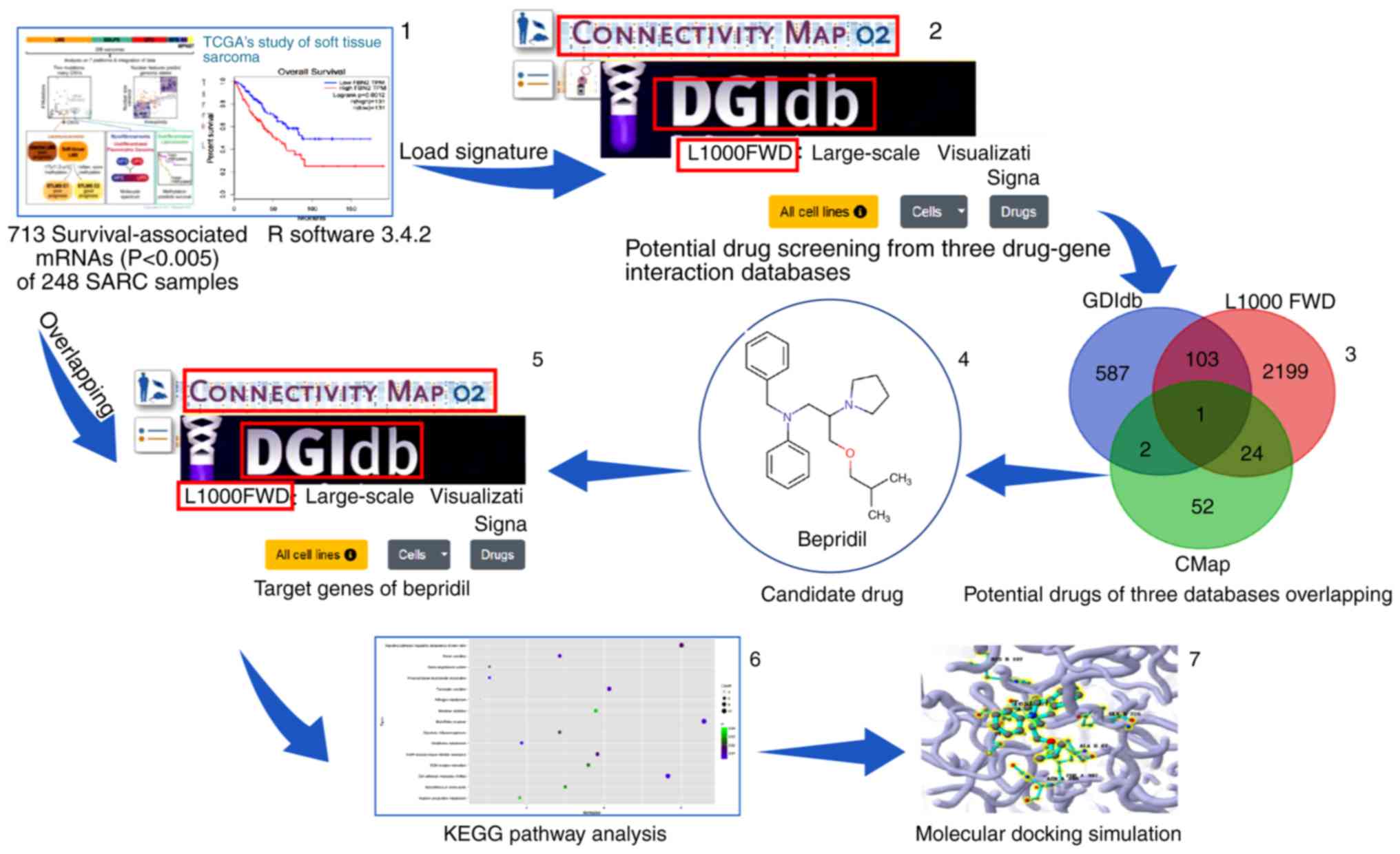 Novel Drug Candidate For The Treatment Of Several Soft
 Storing Genetic Information Biology For Majors I
 A Web Tool For Generating High Quality Machine Readable
 Stages Of Translation Article Khan Academy
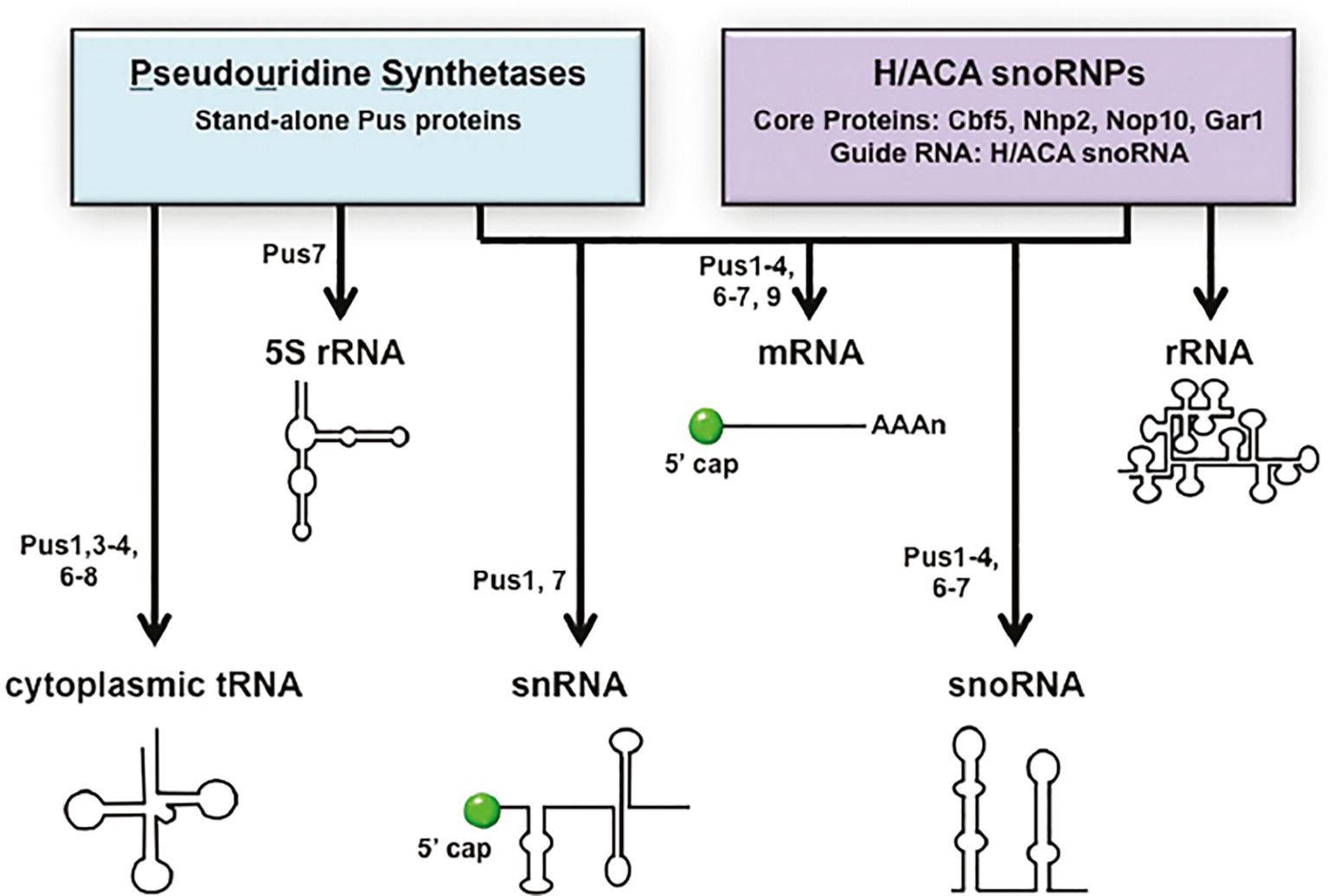 Frontiers Trna Processing And Subcellular Trafficking
 The Signal Peptide Plus A Cluster Of Positive Charges In

 Iso Seq Allows Genome Independent Transcriptome Profiling Of
 Tandem Subunits Effectively Constrain Gabaa Receptor
Belum ada Komentar untuk "Which Protein Subunits Are Depicted In The Diagram"
Posting Komentar