Eye To Brain Connection Diagram
How the brain works with the eyes. Wiring diagram of retinal neurons is first step toward mapping the human brain.

Try our crash course in eye anatomy.

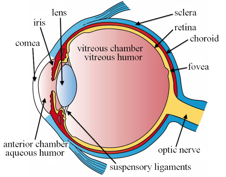
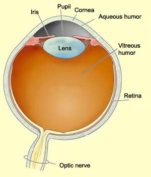
Eye to brain connection diagram. The cornea pupil lens iris retina and sclera. The optic nerve is mainly composed of retinal ganglion cell rgc axons. Making connections in the eye.
Labeled diagram of the eye. The dark circular opening in the center of the iris of the eye varying in size to regulate the amount of light reaching the retina. These impulses are then sent through the optic nerve and to the brain.
This helps us to understand how each one is situated and related to the other. On a diagram of the eye we can see all of the relevant structures together on one image. Through the optic nerve and through the cerebral cortex at the stem of the brain the electrical impulses have officially left the eye.
Eye brain connection experiment with pilots. Multiple genes inherited from each parent determine a persons eye color. 950 neurons reconstructed in a block of mouse retina imaged using serial block face electron microscopy gray images.
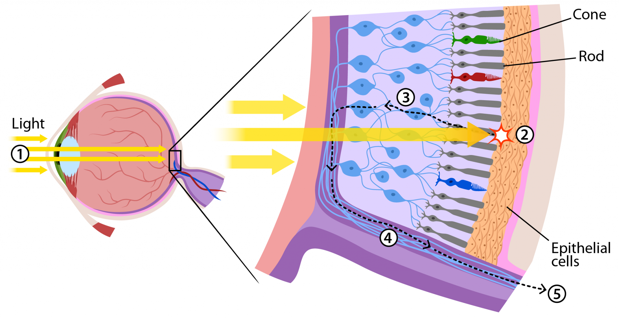
In addition such forms of treatment are held by favoring the myth that bad vision cannot be corrected. Spheres indicate cell bodies red ganglion cells green amacrine cells. Once these rods and cones receive the images they are converted into electrical impulses.
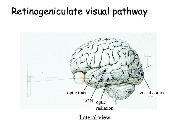
These work together to capture an image and transmit it directly to the brains occipital lobe via the. Start studying eye anatomy and function. The optic nerve a cablelike grouping of nerve fibers connects and transmits visual information from the eye to the brain.
The macula is a small extra sensitive area in the retina that gives you central vision. Change your vision change your brain change your life. Behind the eye your optic nerve carries these impulses to the brain.
Treatment of symptoms by surgery eyeglasses and contact lenses can only temporarily help people see better but shortly afterwards comes to a weakening of eyesight. Eye color is created by the amount and type of pigment in your iris. Learn vocabulary terms and more with flashcards games and other study tools.
One of our favorite ways to get to grips with all of the parts of the eye is by utilizing labeled diagrams. The eye has several major components. In the human eye the optic nerve receives light signals from about 125 million photoreceptor cells known as rods and cones via two intermediate neuron types bipolar and amacrine cells.
The colorful structure of the eye that changes the shape of the pupil to allow for different amounts of light to enter the inner eye. Connection of the optic nerve and retina also where the blood vessels.
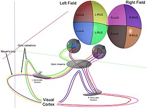
 Visual And Biological Pathways In The Brain Nerve
 Alzheimer S Disease And Glaucoma Is There A Connection
 Visual System Wikipedia
 The Brain And Spinal Cord Canadian Cancer Society
 Visual Perception More Than Meets The Eye Answers In
 Visual Processing Cortical Pathways Section 2 Chapter 15
 Biology Eye Diagram Label Schematic Wiring Diagram
 Primary Projection Areas For Vision Bio Bases Brain

 Perception Lecture Notes Lgn And V1


 How The Spinal Cord Works Reeve Foundation
 Neural Pathway Wikipedia
 How Eye Work Medical Illustration Eye Brain Diagram Canvas Print
 The Eye Is Our Window To The Brain And There S A Lot We
 How Do We See Light Ask A Biologist
 The Eye Brain Connection Health Brain Connections Brain
 Connection Between Eyes And Brain Diagram Quizlet
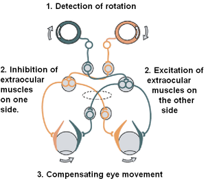 Vestibulo Ocular Reflex Wikipedia
![]() Human Eye Images Stock Photos Vectors Shutterstock
Human Eye Images Stock Photos Vectors Shutterstock
 Diagram Of A Eye Catalogue Of Schemas
How Alzheimer S Disease Affects Vision And Perception
 Structure And Function Of The Eyes Eye Disorders Merck
 Human Eye Ball Anatomy Physiology Diagram
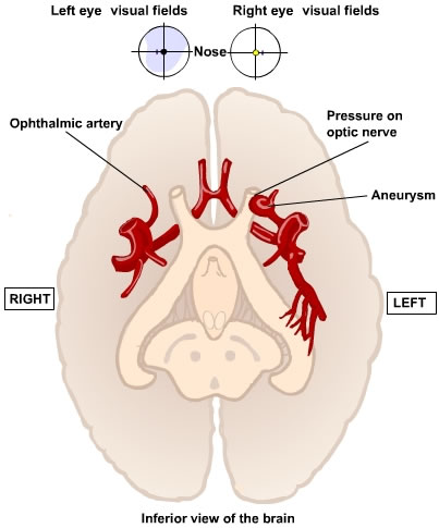
 Which Side Of The Brain Does The Optic Nerve Connect To
 The Brain From Top To Bottom
Belum ada Komentar untuk "Eye To Brain Connection Diagram"
Posting Komentar