Diagram Of Uterus And Bladder
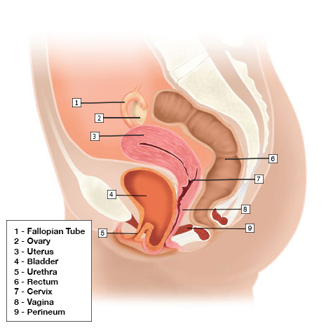
The fundus lies above the entrance of the uterine tube. The bladder is lined by layers of muscle tissue that stretch to hold urine.

The actual position of the uterus within the pelvis varies from person to person.

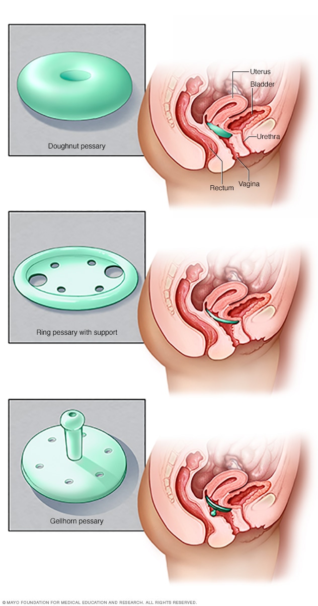
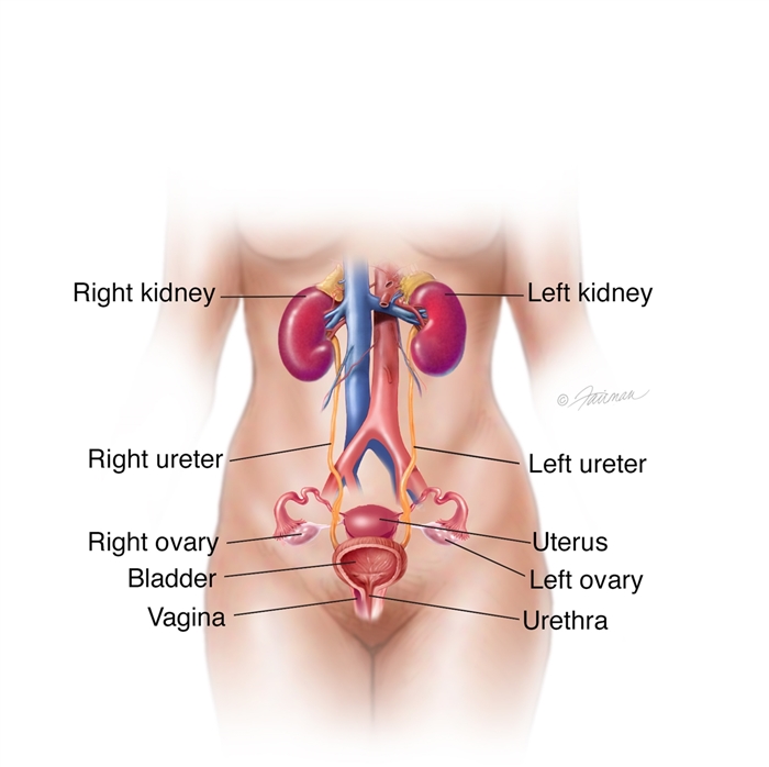
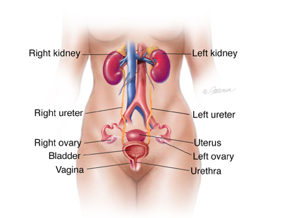
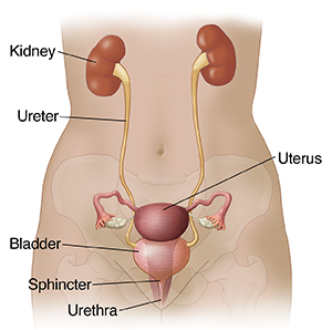
Diagram of uterus and bladder. The uterus is a pear shaped hollow organ with muscular walls. When empty the bladder is about the size and shape of a pear. The normal capacity of the bladder is 400 600 ml.
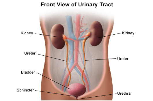
During urination the bladder muscles squeeze and two sphincters. Each organ is connected to the outside by a tube urethra which is about 4cm long vagina which is 10 12cm long and anus also about 4 cm long. Diagrams of uterine and vaginal prolapses rectocele diagram surgery female genital anatomy images peritoneal body cavity uterus bladder anterior wall urethra opening cervix diagram of fetus in uterus overview the is an organ female reproductive system location sits middle pelvis.
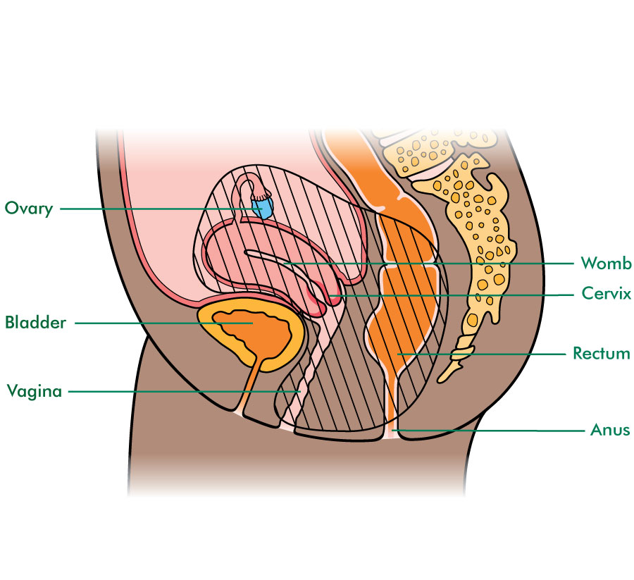
Bladder bowel and uterus. The bladder is a musculomembranous sac located on the floor of the pelvic cavity anterior to the uterus and upper vagnia in females. In a nonpregnant female it lies on the urinary bladder.
Posted on march 25 2019 by admin. The inner lining of the bladder tucks into the folds and expands out to accommodate liquid. The liver stomach and abdominal contents are clearly identified and labeled including the cecum ascending colon transverse colon descending colon and small intestine.
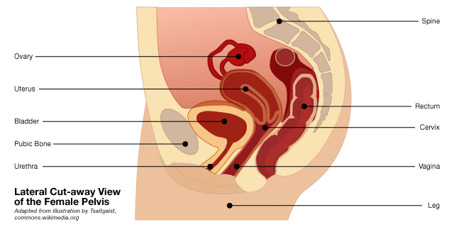
The female pelvic organs. Its function is to nourish a fertilized ovum. The image also shows the pelvis uterus and urinary.
The body is the part below. The blood from your periods and your baby also pass through the vagina. In the bladder diagram above you can see the structure of the urinary bladder.
This medical exhibit diagram illustrates the anatomy of the female abdomen and pelvis from an anterior front cut away view showing elements of the digestive system. The cervix is the narrow part that protrudes into the vagina. The bladder stores your urine the uterus your baby and the rectum your faeces.
78085441 fistula between bladder and uterus or vagina and rectum and uterus. Bladder vagina uterus fallopian tube ovaries. The upper and side surfaces of the bladder are covered by peritoneum also called serosa.
The uterus sits in the middle of the pelvis behind the bladder and in front of the rectum. Bladder diagram uterus image via humanbodyanatomyco. Outer surfaces of the bladder.
The uterus and the vagina.
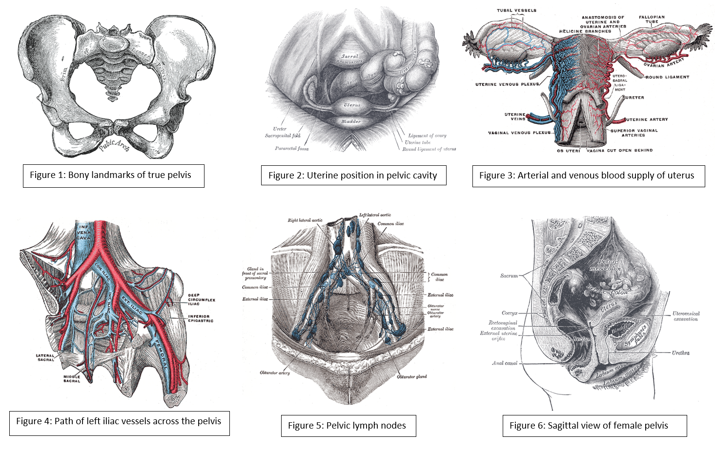
 Uterine Prolapse Diagnosis And Treatment Mayo Clinic
 What Is Pelvic Exenteration In Women Information And

Uterine Vaginal Prolapse Cleveland Clinic
 This Medical Illustration Features An Anterior View Of The
 Vesicovaginal Fistula Wikipedia
 The Vagina Vulva Female Anatomy Pictures Parts
 Kidney Stones Urology Care Foundation
![]() Pelvic Rehab Therapy Help For Uncomfortable Postpartum
Pelvic Rehab Therapy Help For Uncomfortable Postpartum
 Vesicoureteral Reflux Vur Niddk
 Nerve Distribution Of The Bladder And Uterus Medical


 Uterine Fibroid Pictures Anatomy Diagrams Pictures Of
 What Is Nocturia Urology Care Foundation
 Cystocele Austin Urogynecology
 Prolapsed Bladder Signs Symptoms What To Do Always
 Urinary Bladder Wikipedia
 Rectocele Diagram Surgery Female Genital Anatomy Images

 Asymptomatic Uterine Incarceration At Term Successful
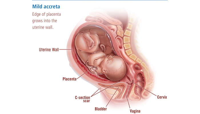 Placenta Accreta Overview Brigham And Women S Hospital
 Bladder Cancer Treatment Bladder Cancer Pictures Signs
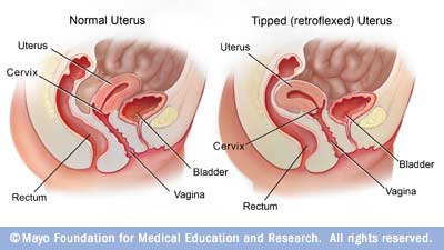 What Is A Tilted Uterus And When Will It Flip Forward
Belum ada Komentar untuk "Diagram Of Uterus And Bladder"
Posting Komentar